The Photon Surgeons: How Controlled Light Is Rendering the Scalpel Obsolete
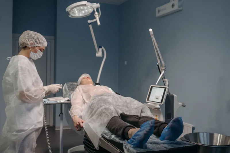
For most of modern medicine's history, surgery has been an act of controlled damage. To remove a tumor, a surgeon must cut through healthy tissue. To stop bleeding, a cauterizing instrument burns. The operating room, for all its technological sophistication, has remained a place where healing begins with deliberate injury. That premise is now under serious challenge — not from robotics or artificial intelligence alone, but from light itself.
Across research hospitals in Boston, Houston, San Francisco, and Ann Arbor, a new generation of photonic tools is demonstrating that beams of precisely tuned light can accomplish what a blade cannot: cutting at the cellular level without mechanical trauma, diagnosing malignancies in real time, and activating therapeutic agents with pinpoint spatial control. The convergence of these capabilities, researchers argue, could fundamentally alter the surgical suite before 2030.
What Happens When Light Meets Living Tissue
The interaction between photons and biological matter is neither simple nor uniform. Different wavelengths penetrate tissue to different depths, scatter at different rates, and are absorbed by different molecular targets. Hemoglobin, water, melanin, and lipids each respond to specific spectral windows — a fact that photonic medicine exploits with growing sophistication.
Near-infrared light, for instance, passes through several centimeters of tissue with relatively little absorption, making it useful for imaging structures beneath the skin. Ultraviolet wavelengths, by contrast, are absorbed almost immediately upon contact with biological molecules, enabling precise surface-level ablation. Carbon dioxide lasers, which operate in the mid-infrared range, are absorbed almost entirely by water — and since soft tissue is largely water, they cut cleanly and efficiently, with minimal thermal spread to surrounding cells.
This spectral specificity is the foundational logic of photonic surgery. Rather than relying on a blade's mechanical edge, light-based tools exploit the physics of absorption to achieve effects that are, in many respects, more controllable than any scalpel.
Photodynamic Therapy's Expanding Frontier
Among the most clinically mature photonic approaches is photodynamic therapy, or PDT. The technique involves administering a photosensitizing compound — a molecule that remains inert until activated by light of a specific wavelength — which preferentially accumulates in tumor cells. When a clinician directs the appropriate light into the target region, the sensitizer generates reactive oxygen species that destroy the malignant tissue from within, leaving adjacent healthy structures largely unaffected.
The National Cancer Institute has supported PDT research for decades, and the approach is already approved for treating certain esophageal and lung cancers, as well as specific dermatological conditions. But the frontier has moved well beyond these initial applications. Researchers at the University of Texas MD Anderson Cancer Center and at Northwestern University's Feinberg School of Medicine are investigating next-generation photosensitizers that are activated by near-infrared light — enabling treatment of deeper tumors that earlier generations of PDT could not reach.
Simultaneously, nanoparticle delivery systems are being engineered to carry photosensitizing agents directly to tumor sites with greater specificity, reducing the systemic exposure that has historically limited the technique's tolerability. The combination of improved sensitizers, deeper-penetrating wavelengths, and targeted delivery represents what many in the field describe as PDT's second era.
Optical Coherence Tomography: Seeing Without Cutting
If PDT represents the therapeutic arm of photonic medicine, optical coherence tomography — OCT — is its diagnostic counterpart. The technology uses low-coherence interferometry to produce cross-sectional images of tissue microstructure at resolutions approaching ten micrometers, roughly comparable to conventional histology. In practical terms, it allows clinicians to see inside tissue without removing it.
OCT is already standard in ophthalmology, where it has transformed the diagnosis and monitoring of retinal disease. Its expansion into cardiology, gastroenterology, and surgical oncology is now accelerating. Researchers at Massachusetts General Hospital and MIT's Research Laboratory of Electronics have collaborated on intraoperative OCT systems that give surgeons real-time subsurface visualization during procedures — effectively providing a live biopsy-quality view of the operative field without requiring tissue excision.
The implications for cancer surgery are substantial. One of the persistent challenges in tumor resection is ensuring that margins are clear — that no malignant cells remain at the edges of the removed tissue. Currently, this verification requires sending samples to a pathology laboratory, a process that can take days. Intraoperative OCT, proponents argue, could compress that feedback loop to seconds, allowing surgeons to confirm clean margins before the patient leaves the table.
Laser Ablation and the Precision Imperative
Beyond PDT and OCT, laser ablation technologies are being refined for applications ranging from neurosurgery to orthopedics. Laser interstitial thermal therapy, or LITT, uses a laser fiber inserted through a small burr hole in the skull to ablate brain tumors and epileptic foci with millimeter-level precision. The procedure, guided by real-time MRI thermometry, allows neurosurgeons to destroy target tissue while preserving eloquent brain regions that would be at risk in open surgery.
Several American medical device companies — including Visualase, now owned by Medtronic, and Monteris Medical — have received FDA clearance for LITT systems, and the procedure is now performed at major academic medical centers across the country. Clinical data from centers including the Cleveland Clinic and Johns Hopkins suggest that LITT can achieve outcomes comparable to open resection for select tumor types, with significantly reduced recovery times and hospital stays.
In orthopedics, cold atmospheric plasma and ultrashort-pulse laser systems are being investigated for cartilage reshaping and bone cutting with thermal damage profiles far narrower than conventional surgical lasers. The goal is a tool that operates with the precision of a focused beam while producing the biological response of a gentle incision.
The Regulatory and Integration Challenge
For all their promise, photonic surgical technologies face a demanding path through the American regulatory landscape. The FDA's Center for Devices and Radiological Health evaluates these systems under frameworks that were largely developed for mechanical and electrosurgical instruments — frameworks that do not always map cleanly onto the novel risk profiles of photonic devices.
The challenge is compounded by the speed of innovation. Photosensitizer formulations, nanoparticle delivery platforms, and AI-assisted guidance systems are each evolving rapidly, and the combination of these elements into integrated therapeutic systems raises questions about how to assess safety and efficacy holistically rather than component by component.
Researchers and industry representatives have called for updated regulatory guidance that accounts for the convergent nature of modern photonic medicine — guidance that acknowledges, for instance, that the safety profile of a PDT system depends not only on the light source but on the sensitizer, the delivery mechanism, and the imaging platform used to confirm dosimetry.
Toward the Photonic Operating Room
What might a photon-powered surgical suite look like by 2030? Researchers sketching that future envision a space in which multiple light-based modalities operate in concert: an OCT system providing continuous subsurface visualization, a laser ablation platform guided by AI-assisted image analysis, and a PDT delivery system triggered by real-time feedback from a spectroscopic sensor monitoring tissue response.
Such integration is not speculative engineering. Its components exist today, in various stages of clinical validation, distributed across American research institutions and device companies working in parallel toward a common convergence point. The question is less whether these technologies will reach the operating room than how quickly the systems-level work — the integration, the regulatory clearance, the clinical training — can be completed.
For the patients who will benefit, the difference may be measured not in years of survival but in weeks of recovery, in the absence of surgical scars, in the preservation of function that a mechanical approach would have sacrificed. That, in the language of photonic medicine, is what precision actually means.